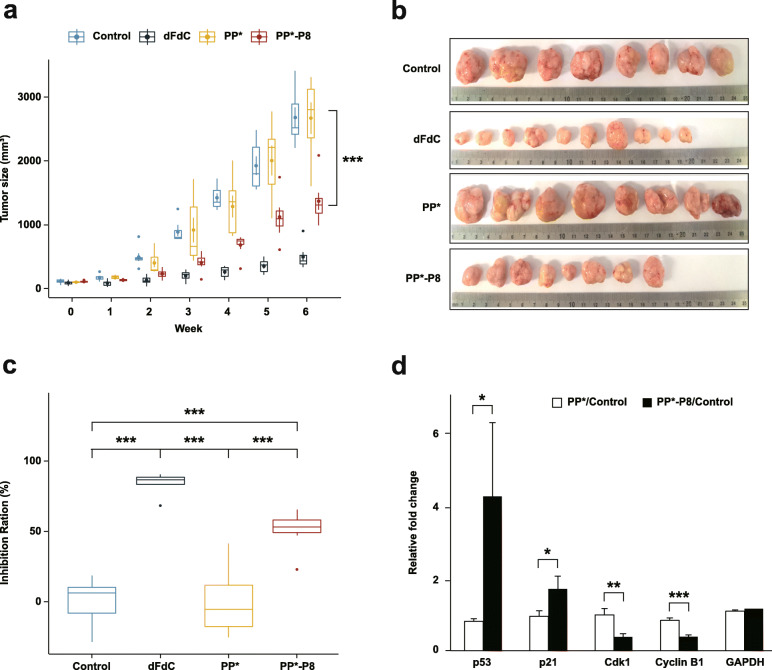
Fig. 2.
Anti-tumor efficacy of the PP*-P8 probiotic in the DLD-1 xenograft mouse model. a Increased sizes of DLD-1-derived tumors recorded each week. Mice (n = 10 in each group) were subcutaneously inoculated with 2 × 10⁶ DLD-1 cells in the rear right flank and then received 0.9% saline (control), 60 mg/kg body weight gemcitabine (dFdC; intraperitoneal injection, twice a week), 1 × 10¹⁰ CFU/head P. pentosaceus alr (pCBT24-2-alr) (PP*; oral administration, five times a week), or 1 × 10¹⁰ CFU/head P. pentosaceus alr (pCBT24-2-PK-p8-PK-p8-alr) (PP*-P8; oral administration, five times a week). ***P < 0.001 for control vs. dFdC, control vs. PP*-P8, dFdC vs. PP*, PP* vs. PP*-P8. b Extracted tumor tissues from each treatment group 6 weeks after the DLD-1 xenograft. c Inhibition ratios for tumor growth calculated from the mean tumor weights of the control group and the test groups. ***P < 0.001. d Relative fold changes in the expression of cell cycle regulatory factors between PP* with control and PP*-P8 with control. Each vertical bar represents the arithmetic mean of three replicates. *P < 0.05, **P < 0.01, ***P < 0.001

