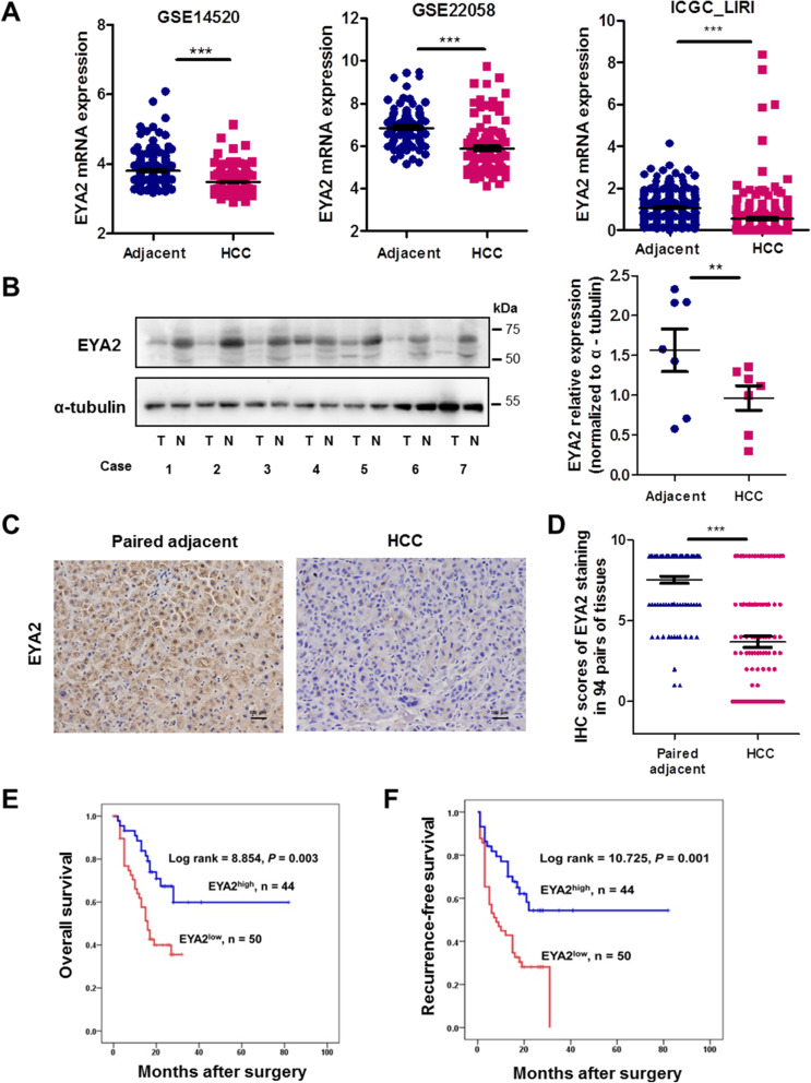
Fig. 2.
Expression and prognostic significance of EYA2 in HCC tissues. (A) EYA2 expression in HCC and adjacent tissues according to the three datasets GES22058, GSE14520 and ICGC_LIRI. (B) Western blot analysis of EYA2 expression in HCC and adjacent tissues. T: HCC tissues, N: Adjacent tissues. (C) A representative immunohistochemical staining of EYA2 in HCC and case-matched adjacent tissue. (D) Immunohistochemistry scores associated to the EYA2 expression in HCC and case-matched adjacent tissues (n = 94 pairs). Kaplan-Meier analysis of overall survival (E) and recurrence-free survival (F) of HCC patients with high or low EYA2 expression (n = 94 cases). **P < 0.01, ***P < 0.001. (A, B, D) mean ± SEM, Student’s t-tests; (E, F) Kaplan-Meier analysis

