Fig. 4.

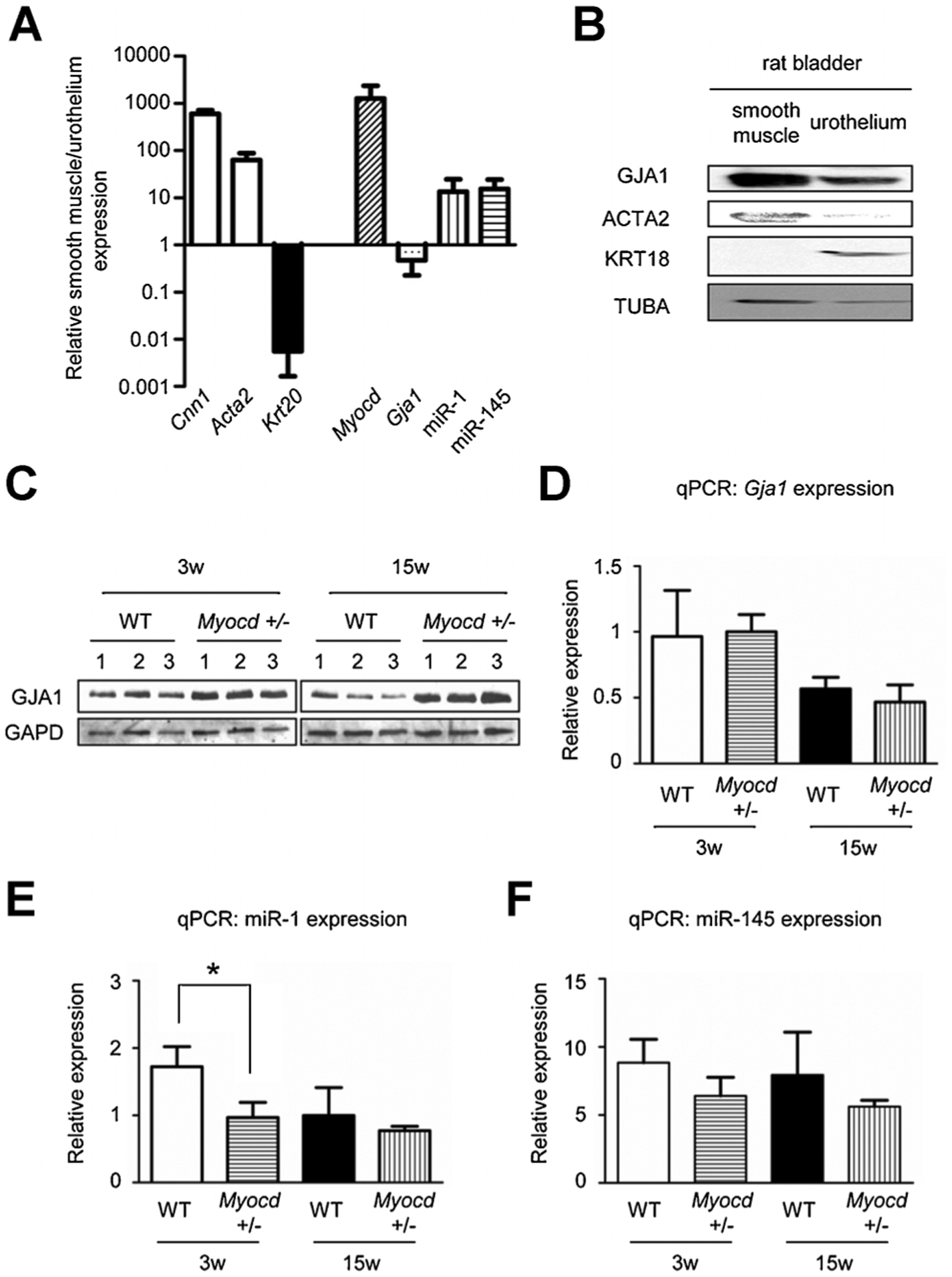
A: qPCR data showed that Myocd and miR-1 was predominantly expressed in the smooth muscle layers which was positive for smooth muscle markers (Cnn1, Acta2) but not in urothelium which was positive for an urothelial marker (Krt20). Gja1 was expressed in both. B: Western blotting showed that GJA1 protein was predominantly expressed in the smooth muscle layers positive for a smooth muscle marker (ACTA2) but not in urothelium positive for urothelial marker (KRT18). C: Western blotting revealed up-regulated protein level of GJA1 in bladders of 3 and 15-week-old Myocd+/− mice compared to those of WT mice. D: In contrast, qPCR data showed no difference in RNA level of Gja1 in bladders of WT and Myocd+/− mice. E: qPCR data showed miR-1 in bladders of Myocd+/− mice was down-regulated compared to those of WT mice. F: qPCR data also showed miR-145 in bladders of Myocd+/− mice was down-regulated compared to those of WT mice. *Statistically significant difference (P < 0.05).
