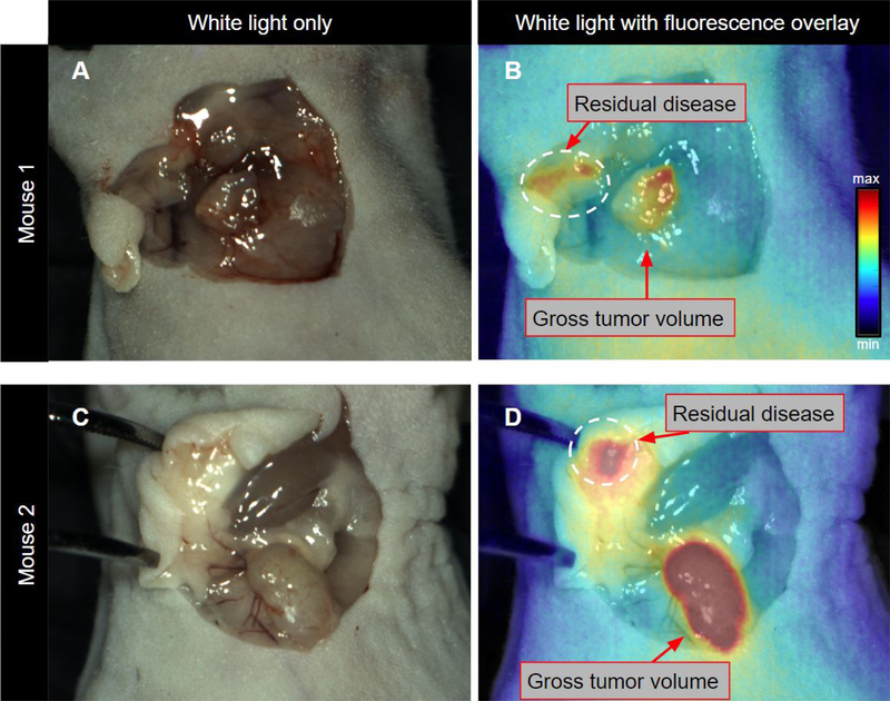
Figure 2. Intraoperative visualization of residual disease following gross tumor resection under ambient light.

White light in situ visualization after tumor resection using direct visual inspection and palpation only (for the proof-of-concept study tumors were placed back in the field of view) (A and C). Tumor beds were then surveyed using the OnLume FGS imaging system and residual fluorescence was detected (circumscribed by dashed white circle) (B and D). B and D are the corresponding NIRF images overlaid on A and B, respectively.
