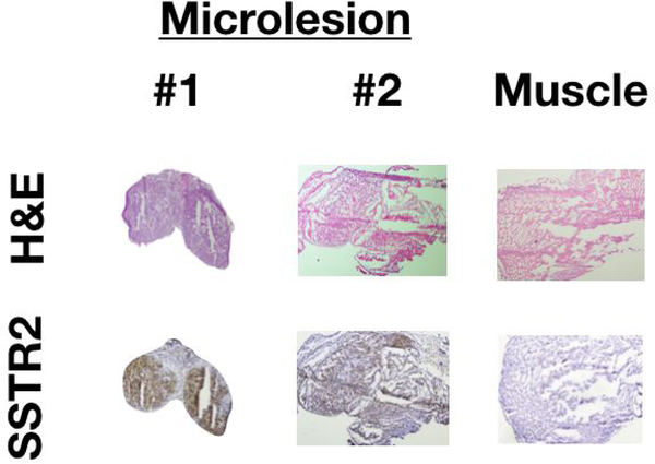
Figure 4. Histological analysis of residual fluorescence was confirmed for cancer positivity and SSTR2-overexpression.
The top row shows H&E staining for tumor and muscle (n = 2). The bottom row shows corresponding IHC staining, which indicates SSTR2 presence only in tumors (malignant cells: brown areas).

