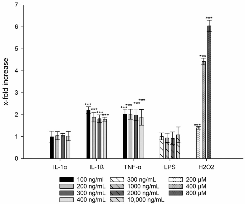
Figure 3.
Generation of ROS after treatment with IL-1α, IL-1ß, LPS, and TNF-α compared to untreated cells. H2O2 was used as positive control. Student´s t-test was used for statistical analysis. Significant differences compared to the control are marked as ***, which corresponds to a p value ≤ 0.001.

