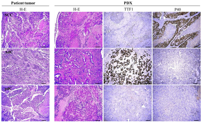
Figure 1.
Histological evaluation of primary tumors and PDX. Images from eosin and hematoxylin staining (H/E) from a representative example of squamous cell carcinoma (SCC), adenocarcinoma (AdC), and a poorly differentiated carcinoma (pdC) showing that the mouse tumor preserved the morphological features of the primary tumor. The squamous cell marker P40 and the adenocarcinoma marker TTF1 were also used on the PDX tumor. Scale bar: 100 μm.

