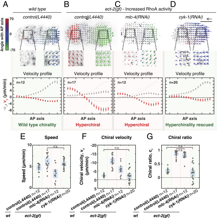
Fig. 4.
RhoA promotes chiral counterrotating actomyosin flow via CYK-1/Formin activation. (A–D) (Top) Time-averaged flow fields overlaid on a still image of cortical NMY-2::GFP of (A) a wild-type embryo on RNAi control (L4440), (B) an ect-2(gf) mutant on RNAi control (L4440), (C) ect-2(gf); mlc-4(RNAi), and (D) ect-2(gf); cyk-1(RNAi). Mean velocity vectors are color coded for their angle with the anteroposterior axis. (Scale bar, 10 m.) Velocity scale arrow, 20 m/min. (Bottom) Mean x velocity (gray) and y velocity (red) in 18 bins along the anteroposterior axis. Dashed lines in the plots in B–D display the mean x- and y-velocity profiles in wild type. Error bars, SEM. n indicates the number of embryos. (E–G) Mean speed (E), chiral velocity (F), and chiral ratio (G) per embryo. Data points represent individual embryos. Blue stripe and area represent the mean with 95 confidence interval. Significance testing: Only conditions that are not significantly different are indicated in the diagrams (n.s., P 0.05, Wilcoxon rank sum test). The flow hyperchirality (red background in all panels) of ect-2(gf) is rescued in ect-2(gf); cyk-1(RNAi) (green background in all panels), but not in ect-2(gf); mlc-4(RNAi) (red background in all panels).

