Fig. 5.
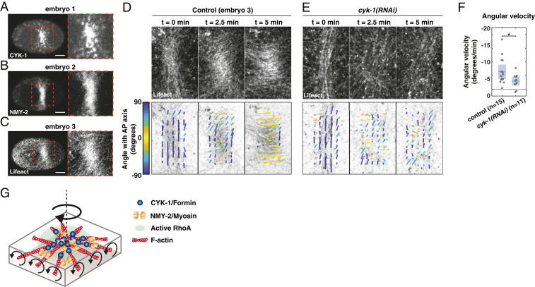
CYK-1/Formin activity in a compression-induced RhoA patch promotes clockwise reorientation of cortical F-actin. (A–C) Micrographs of compressed embryos in which the cytokinetic ring collapsed, resulting in a region enriched in (A) CYK-1/Formin, (B) NMY-2, and (C) F-actin. Note that the three images are derived from three different embryos. (Scale bars, 10 m.) (D and E) Fluorescent micrographs of cortical Lifeact-mKate2 (Top), overlaid with the local filament order in small templates (Bottom) of a (D) control and (E) cyk-1(RNAi) embryo, at the onset of patch formation (t = 0 min), during patch rotation (t = 2.5 min), and at the end of patch rotation (t = 5 min). Both examples shown in D and E display an embryo with strong clockwise reorientation, relative to the mean of the condition. Local directors are color coded for the angle with the anteroposterior axis. (F) Mean angular velocity in a time window of 5 min during reorientation of the patch in control and cyk-1(RNAi). Because the timing of rotation with respect to the onset of patch formation varied among embryos, the start of the 5-min time window was chosen such that the clockwise angular velocity was maximal (Materials and Methods). Mean with 95 confidence interval is indicated in blue. Significance testing: *P 0.05 (Wilcoxon rank sum test). n indicates the number of embryos. (G) Schematic of an active RhoA patch in which F-actin, NMY-2, and CYK-1/Formin are enriched. If Formins in the RhoA patch are constrained and elongate filaments in opposite directions, opposing filaments will counterrotate, which we hypothesize drives clockwise rotation of the patch as a whole.

