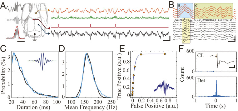
Fig. 5.
MTA-based neural implant enables real-time closed-loop suppression of hippocampal ripples. (A) Schematic of anatomical location of recording (HC = hippocampus, blue; mPFC, orange) and stimulation electrodes for ripple suppression (Left). The sample raw traces from real-time simultaneous recording showing hippocampal signal (black trace), hippocampal signal processed for detection (green), and stimulation triggers (red) during NREM sleep (Scale bar, 100 ms, 500 µV). (B) Sample traces from MTA-device recording showing physiologic co-occurrence hippocampal ripple (ρ), cortical delta (δ), and sleep spindle (σ) with visible action potentials. (Scale bar, 100 ms, 500 µV). (C) Comparison of detected ripple duration obtained by the real-time closed-loop MTA-based system (blue) with offline detection of ripples obtained by conventional neural acquisition system (black) within the same animal (n = 990). (D) Comparison of detected ripple mean frequency obtained by the real-time closed-loop MTA-based system (blue) and offline detection of ripples obtained by conventional neural acquisition system (black) within the same animal (n = 990). (E) Receiver operating characteristics curve for the online ripple detector (n = 1,935). (F) Cross-correlogram of online detected ripples and offline detected ripples in closed-loop suppression mode (CL; n = 2,266) and detection-only mode (Det; n = 1,061). Inset: Sample of a closed-loop stimulation triggered by a ripple (black trace = LFP in hippocampal CA1 pyramidal layer; orange box = ripple oscillation cycles triggering detection [Scale bar, 100 ms, 200 µV]).

