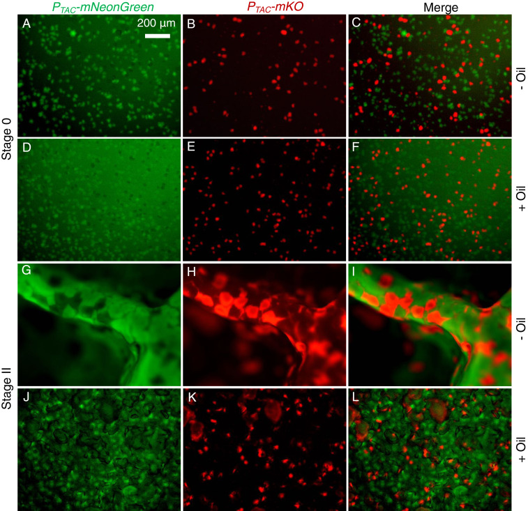
Fig. 4.
Individual V. cholerae microcolonies remain segregated, and the microstructural “memory” of the pellicle is preserved despite global morphological transitions. A 1:1 mixture of cells of two otherwise isogenic V. cholerae strains constitutively expressing either mNeonGreen (green) or mKO (red) was used to inoculate pellicles. The combined initial inoculum is OD600 = 0.01. (A–F) Top views of the distributions of V. cholerae pellicle microcolonies in Stage 0 for (A–C) a liquid–air interface (no mineral oil) and (D–F) a liquid–liquid interface (with mineral oil). The left column shows colonies expressing PTAC-mNeonGreen, the middle column shows colonies expressing PTAC-mKO, and the right column shows the merged images. (G–L) Top views of the distributions of V. cholerae pellicle microcolonies during Stage II secondary ridge instabilities for (G–I) at the liquid–air interface (no mineral oil) and (J–L) at the liquid–liquid interface (with mineral oil). (Scale bar, 200 μm.)

