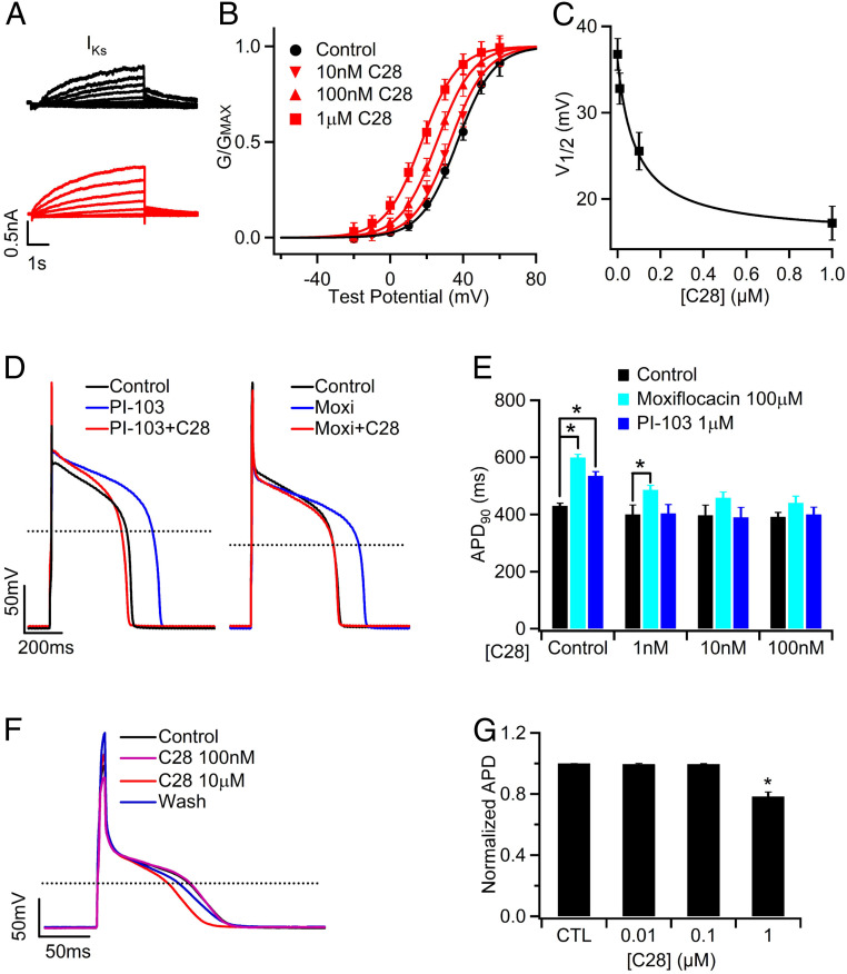
Fig. 6.
C28 enhances IKs and stabilizes APs in GP ventricular and atrial myocytes. (A) IKs currents, measured as the Chromanol 293B sensitive currents (SI Appendix, Fig. S8 A and B), of control (black) and with C28 (100 nM, red) at various voltages (see B) from a ventricular myocyte. The voltage before and after the test pulses were −40 and −20 mV, respectively. (B) IKs G–V relations in various C28 concentrations. The lines are fits to Boltzmann equation. (C) V1/2 of G–V relations versus C28 concentration. (D) C28 (100 nM) on APs in control and in the presence of PI-103 (1 µM) and Moxifloxacin (Moxi, 100 µM). (E) Effects of C28 on APD. *P < 0.05, n = 5 to 44. (F) APs of a GP atrial myocyte recorded in control, C28 (100 nM and 10 µM), and after washout (Wash). The stimulus was 180 pA in amplitude and 10 ms in duration at 1 Hz frequency. (G) Effects of C28 on atrial APD. *P < 0.05 compared to control, n = 9 to 12.

