Fig. 3.
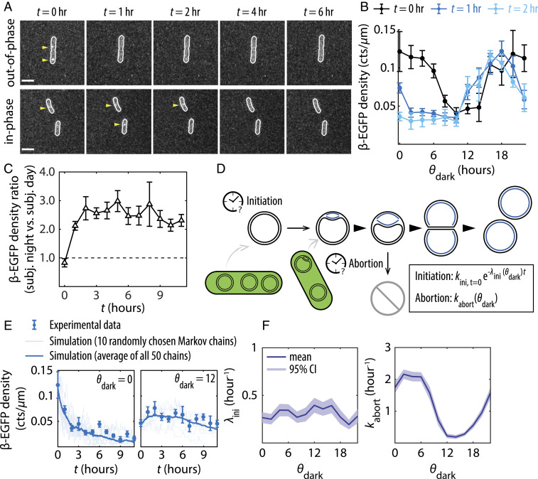
The ability to sustain DNA replication in the dark depends on the clock state at the onset of dark. (A) Fluorescence images showing β-EGFP foci at various time points (t) in the dark, for cells subject to a dark pulse either out-of-phase () or in-phase ( = 12) with respect to their entrainments. (Scale bars = 3 µm.) (B) β-EGFP densities as a function of during the first 2 h of dark (t = 0, 1, 2 h). (C) The ratio of β-EGFP densities between cells in the subjective nighttime state (12 h ≤ < 24 h) and those in the subjective daytime state (0 h ≤ < 12 h) when transferred to the dark, plotted as a function of time in the dark. (D) Schematic diagram of the model used to simulate replication events in the dark. (E) Simulated replication events plotted against experimentally collected β-EGFP data in dark. (F) Best-fit initiation decay rate and best-fit abortion rate plotted as a function of . All β-EGFP error bars are SEMs from five independent experiments totaling ∼27,000 cells.

