Fig. 1.
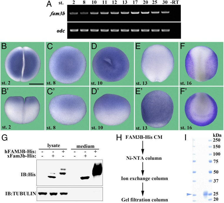
Characterization of the Xenopus fam3b gene and purification of recombinant human FAM3B-His protein. (A) RT-PCR assay showing fam3b expression across different developmental stages. odc was used as a loading control. –RT served as a negative control. (B–F') WISH of fam3b showing its diffuse localization at two-cell (B and B'), blastula (C and C'), and gastrula (D and D') stages (in Xenopus WISH, uniform mRNAs appear stronger in the animal pole due to the accumulation of large yolk platelets in the vegetal pole). At neurula stages, fam3b is localized to the epidermis (E and F'). (Scale bar, 500 μm.) (G) Xenopus fam3b encodes a secreted protein. HEK293T cells were transfected with His-tagged human FAM3B or Xenopus Fam3b constructs. Twenty-four hours after transfection, cells were cultured in serum-free medium for further 20 h. The lysate and medium were subjected to immunoblotting with the indicated antibodies. α-Tubulin served as a loading control and was detected only in the lysate and not in the medium. (H) Flowchart showing purification of His-tagged human FAM3B protein from CM generated from transfected HEK293T cells (Materials and Methods). (I) SDS-PAGE and Coomassie Blue staining showing the purity of recombinant human FAM3B-His protein. Arrowhead indicates the molecular weight of FAM3B protein.

