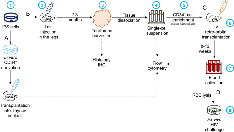
Fig. 1.
Schematic representation of the experimental approaches to assess engraftment of CD34+ cells derived (A) in vitro or (B) in vivo, from iPS cells. (B) 1) iPS cells are expanded in culture before 2) being injected intramuscularly (i.m.) into both hind legs of an NSG mouse; 3) when the teratomas reach 1 cm, the mice are killed and the teratomas harvested for histology and 4) following tissue dissociation, the preparation of a single-cell suspension used for flow cytometry or 5) enrichment of CD34+ cells by immunomagnetic separation. (C) Then, 6) the CD34+ cells are transplanted intravenously (i.v.) into the retro-orbital cavity of irradiated NSG mice; 7) after 6 to 8 wk, the mice are bled to check for engraftment (i.e., presence of HLA-A2+ cells) by flow cytometry. (D) If human cells are detected, 8) these PBMC are used for ex vivo inoculation with HIV isolates to assess HIV sensitivity. RBC, red blood cell.

