Fig. 3.
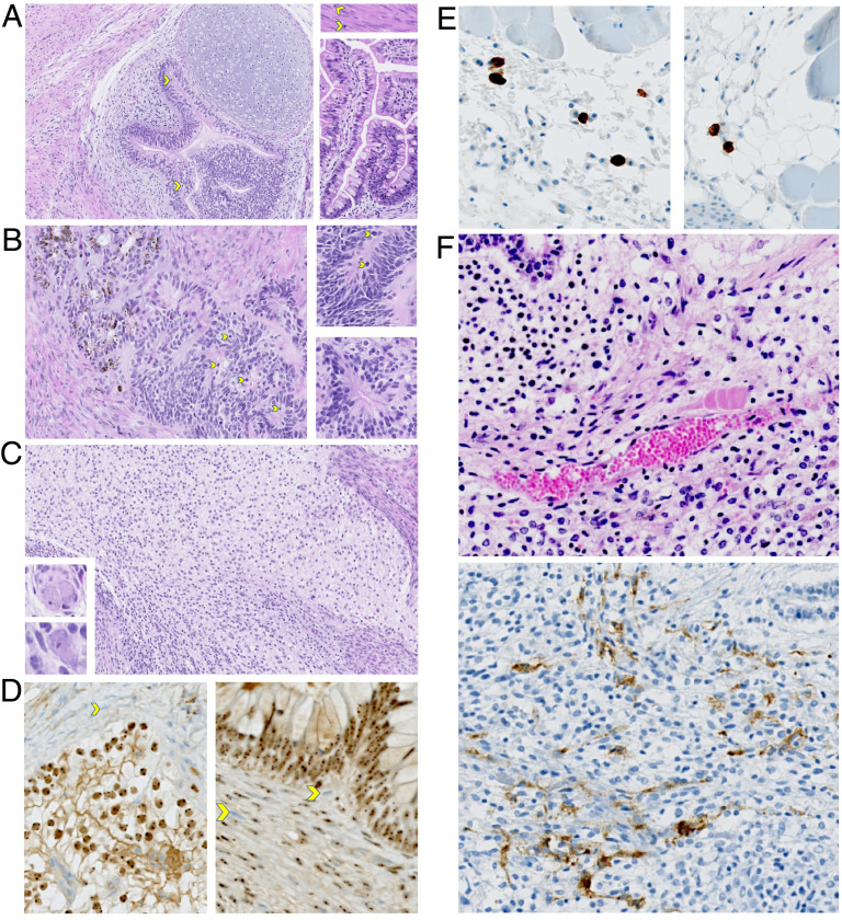
Histologic examination of genetically modified iPS cell-derived teratomas. (A–C) These images demonstrate the typical features of an immature teratoma. (A) The main image shows the pseudostratified neuroepithelium, with mitotic figures (yellow arrows) and overlying cartilage. The Top Right Inset shows smooth muscle with mitotic figures. Bottom Right Inset shows abundant epithelium with goblet cells. Malignant cell populations resembling embryonal carcinoma, choriocarcinoma, and endodermal sinus tumors are completely absent. Magnification, 20×. (B) The main image shows the primitive neuroepithelium with abundant mitotic figures, which is pigmented and resembles early retinal pigment epithelium (RPE), in continuity with nonpigmented neuroepithelium. Both Insets show various types of pseudostratified neuroepithelial rosettes. Magnification, 20×. (C) The main image shows the neuroepithelial-derived neural cell populations that are abundant in this field. Both Lower Left Insets show abnormal ganglionic cells. Magnification, 20×. (D) IHC for human nucleoli was performed. The human cell nuclei are labeled by immuno-peroxidase staining in comparison to the negative mouse cell nuclei showing the blue counterstain in the two images. In the upper field of the Left image, the connective tissue surrounding the immature teratoma is composed of many mouse cells, some indicated by a yellow arrow. In the Right image, mouse cells infiltrating the teratoma are marked by yellow arrows. The microvasculature has both mouse cells and human cells, some of which are CD34+ (illustrated in F). Magnification, 20×. (E) IHC using an antibody specific for human CD3 was performed, labeling human T cells. The two representative images show that CD3+ cells are typically scattered, usually at the mouse–teratoma interface. Magnification, 20×. (F) IHC using an antibody specific for human CD34 was performed. Representative H&E staining (Top) and CD34 staining (Bottom) of fields from the same teratoma are shown. Magnification, 20×.

