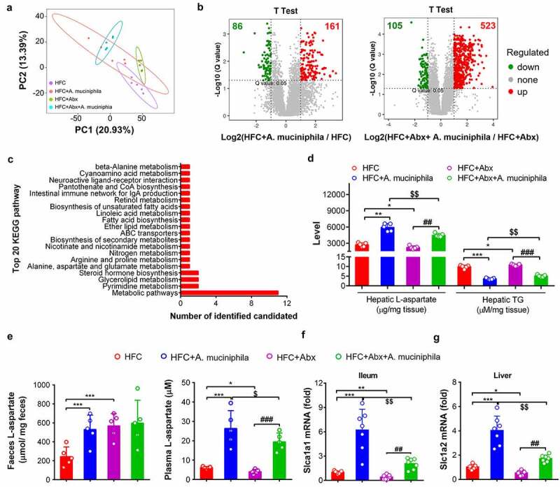
Figure 4.

A. muciniphila increased L-aspartate level in liver of HFC mice. HFC-induced obese MAFLD (11 weeks of feeding) were administered saline (HFC), A. muciniphila, antibiotics (Abx) or a combination of Abx plus A. muciniphila (Abx+A. muciniphila) for (6 weeks of treatment) to assess liver metabolomics and the indicated assays. (a) PCA analysis. (b) Identification of significant differentially metabolites in A. muciniphila- or Abx+A. muciniphila-treated HFC mice compared with HFC or Abx-treated HFC mice by LC-MS analysis. Significant differentially metabolites were identified based on the criteria of fold ≥ 2 and a Q value equal to or higher than 0.05. The dots represent differentially metabolites. N = 5 mice/group. (c) Annotation of the identified significant metabolites in the HFC, HFC+A. muciniphila, HFC+Abx, and HFC+Abx+A. muciniphila groups by metabolomics trend analysis. (d) Comparative analysis of the hepatic L-aspartate and TG levels. (e) L-aspartate level in the feces and plasma of mice. N = 5 mice/group. (f) Gene expression of Slc1a1 in the ileum of mice. (g) Gene expression of Slc1a2 in the liver of mice. The level of mRNA in the HFC control group was set as 1, and relative fold increases were determined by comparison with the HFC control group. N = 5–8 mice/group. * p < .05, ** p < .01, *** p < .001, compared with HFC control mice. # p < .05, ## p < .05, ### p < .001, compared with antibiotics (Abx)-treated HFC mice; $ p < .05, $$ p < .01, $$$ p < .001, compared with A. muciniphila-treated HFC mice
revised Figure 4
