Figure 6.

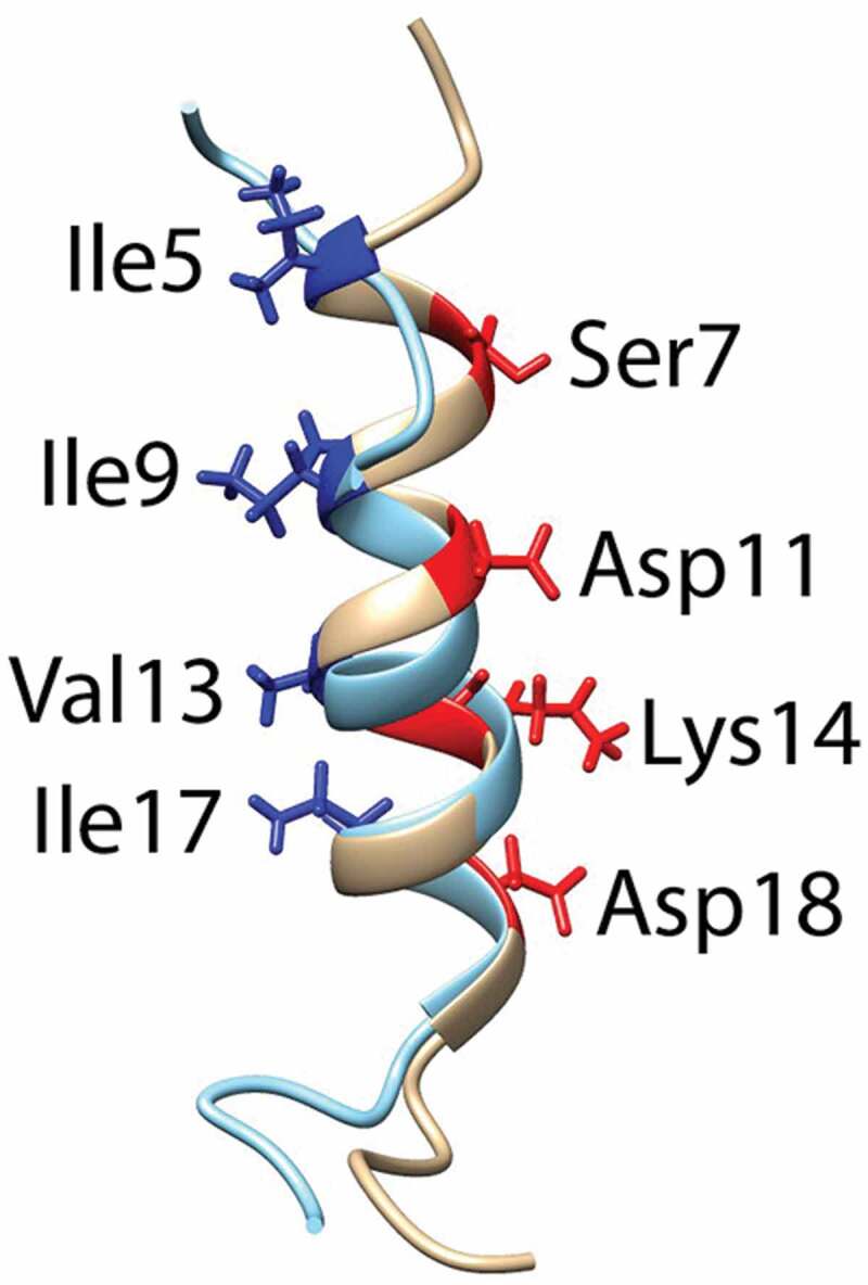
Superimpostion of the NMR structures of dfδ-toxin (gray) and fδ-toxin (cyan) in ribbon mode, with the side chains on the hydrophilic (red) and hydrophobic (blue) sides shown, and the corresponding residues labeled

Superimpostion of the NMR structures of dfδ-toxin (gray) and fδ-toxin (cyan) in ribbon mode, with the side chains on the hydrophilic (red) and hydrophobic (blue) sides shown, and the corresponding residues labeled