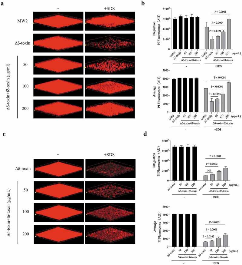
Figure 8.

Impact of adding fδ-toxin fibrils on the structure of static S. aureus biofilms. Static biofilms were grown in eight-well chambered coverglass plates for 48 h, with adding fδ-toxin fibrils (a, b) before the formation of the biofilm (0 h) or (c, d) after the formation of the biofilm (36 h), and to test the dispersal mediated by SDS. (a, c) Three-dimensional confocal laser scanning microscopy (CLSM) images of biofilms. Extensions and scale are the same in every image (total x extension: 160 μm; total y extension: 160 μm). (b, d) Biofilm parameters were measured in at least 5 randomly chosen biofilm CLSM images of the same extension on a Fluoview 1000. Data represent means ± SD. ANOVA was used to determine statistical significance followed by Dunnett’s multiple comparison test. All the experiments were repeated independently three times with similar results
