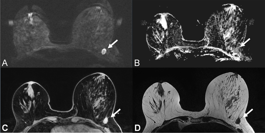
Figure 1:
59 year old patient with a T1c invasive ductal cancer G3. Readout-segmented DWI image at b=850 s/mm2 (A) shows ill-defined strongly hyperintense lesion (arrow) with corresponding low Apparent Diffusion Coefficient values on the ADC map (B). Note the similar contrast and morphologic appearance as compared to the T1w contrast enhanced TWIST image (C) and the T2w-TSE image (D).

