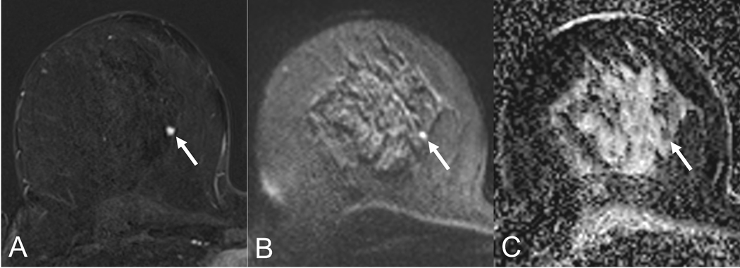
Figure 5:
Lesion conspicuity of a small carcinoma (5mm, arrow) in a 53-year old patient. The lesion shows excellent conspicuity on the early contrast-enhanced subtraction (A) while it is adequately visualized on the b850 s/mm2 image (B). The corresponding ADC map shows a hypointense lesion correlate, corresponding to low ADC-values (0.8 *10−3 mm2/s.

