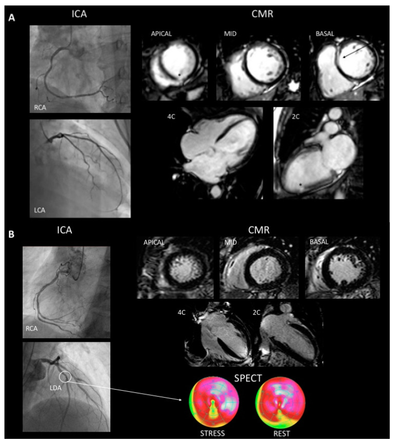
Figure 2.
A 66-year-old woman who presented withg dCMP and bystander myocardial infarction. ICA was normal and CMR showed a 31% LVEF, an end-diastolic volume of 131 mL/m2, global hypokinesia and 2 patterns of LGE. Mid-wall linear septal (arrow) and small sub-endocardial LGE (asterisk) (Panel A). A 71-year-old man who presented dCMP with coronary artery disease. ICA shows plaque to 40–50% on left descending anterior artery (arrow), CMR showed a 32% LVEF, an end-diastolic volume of 123 mL/m2, global hypokinesia and no LGE (Panel B). LCA: Left coronary artery, LDA: left descending artery, RCA: right coronary artery, ICA: invasive coronary angiogram, CMR: cardiac magnetic resonance imaging, SPECT: single photon emission computed tomography.

