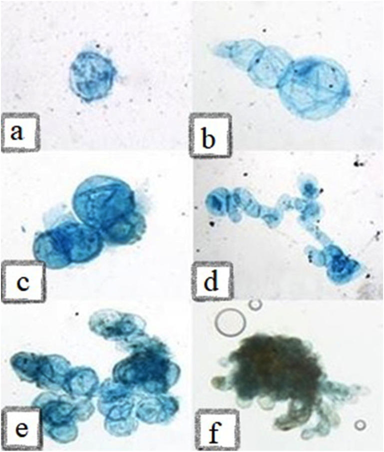
Figure 9. Microscopic illustration of staining cells with Evans Blue (0.1%).
(A) Globular shaped cells with large size, (B) three single interconnected cells, (C-D) starting of cell masses in cell suspension, (E) cell masses with same-sized cells in the presence of 2-4-D, (F) cell masses with elongated cells in the presence of NAA.

