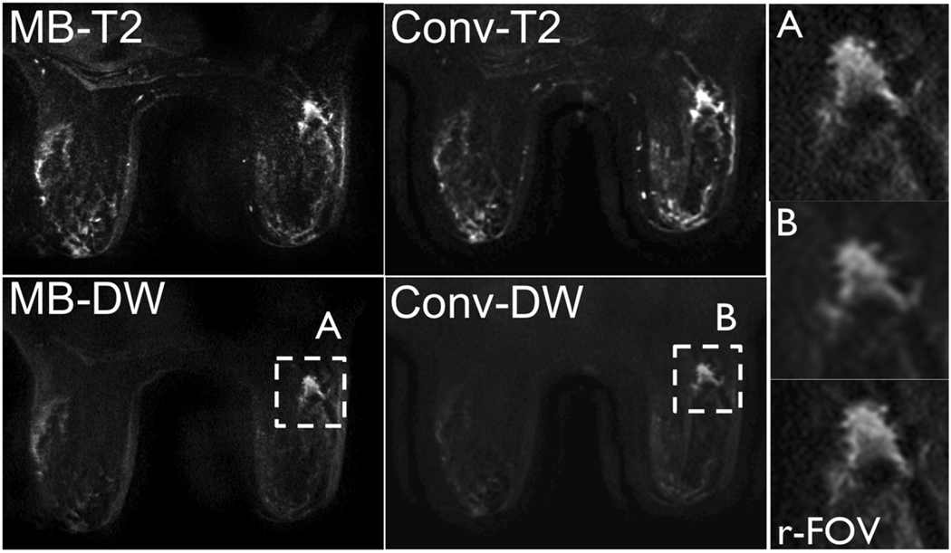
Figure 7.
b = 0 and b = 600s/mm2 images acquired in a 68 year old patient with invasive ductal carcinoma (right breast, 1.6×1.2×1.0cm3) using MB and non-MB DWI. Note the similar distortion field between the MB and non-MB acquisitions and the increased resolution achieved with MB (insert A), similar to that achieved using a targeted reduced-FOV method (r-FOV), with respect to non-MB (insert B).

