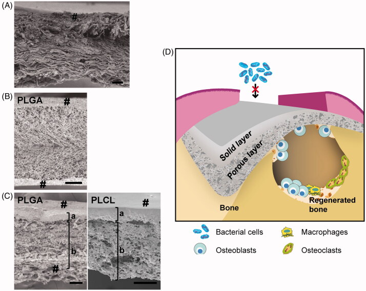
Figure 1.
Cross-sectional images and schematic illustration of GBR membrane. Cross-sectional electron micrographs of (A) collagen membrane (Bio-Gide®), (B) PLGA monolayer membrane and (C) bilayer membranes composed of PLGA (left) and PLCL (right). (D) Schematic illustration of a bilayer membrane for GBR. #: membrane surface; a: solid layer and b: porous layer (b) (scale bar: 100 μm). Reproduced with permission from Yoshimoto et al. [97] and Abe et al. [100]. Copyright Elsevier B.V.

