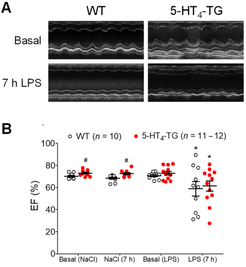
Figure 1.
Echocardiography of LPS-treated mice. (A) M-mode pictures of WT and 5-HT4-TG, basal and 7 h after LPS treatment. (B) LPS treatment (7 h) led to a deterioration of cardiac function demonstrated as decreased left ventricular ejection fraction (EF). Number in brackets indicates the number of mice studied. WT = wild-type mice, 5-HT4-TG=5-HT4-transgenic mice. Data shown are means ± SEM. * p < 0.05 vs. basal; # p < 0.05 vs. WT.

