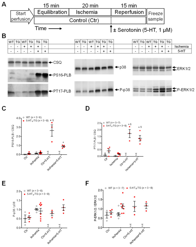
Figure 7.
Protein phosphorylation after ischemia/reperfusion in isolated perfused hearts of WT and 5-HT4-TG mice. (A) The scheme demonstrates the protocols (Langendorff perfusion: 2 mL/min flow): (1) 15 min equilibration, 20 min ischemia by stopping the perfusion followed by 15 min reperfusion or 50 min continuous perfusion with saline buffer as time control; (2) 15 min equilibration, 20 min ischemia and 15 min reperfusion in the presence of 1 µM serotonin (5-HT) or 35 min perfusion followed by 15 min perfusion with 5-HT (1 µM) as time control without ischemia. (B) Representative Western blots. The loading scheme is shown in the table above the blots. TG = 5-HT4-TG. (C) Phosphorylation of phospholamban at serine-16 (PS16-PLB) and (D) threonine-17 (PT17-PLB) normalized to cardiac calsequestrin (CSQ). (E) Phosphorylation of the mitogen-activated protein kinases (MAPK) p38 and (F) ERK1/2 normalized to the non-phosphorylated MAPKs. Ordinates: Ratio of phosphoproteins to calsequestrin or non-phosphorylated MAPKs in arbitrary imager units. Data shown are means ± SEM. * p < 0.05 vs. Ctr; § p < 0.05 vs. ischemia; + p < 0.05 vs. Ctr + 5-HT. WT = wild-type mice, 5-HT4-TG = 5-HT4-transgenic mice; n.d., not determined (As WT preparations did not respond to 5-HT, perfusion with 5-HT was exclusively done with 5-HT4-TG hearts).

