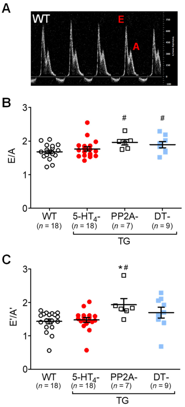
Figure 11.
Doppler echocardiography of double transgenic mice. Pulsed wave (PW) Doppler echocardiography of wild-type (WT), 5-HT4-transgenic (5-HT4-TG), PP2A-transgenic (PP2A-TG) and double transgenic (DT) mice. (A) A typical pattern of E wave and A wave in mitral flow. The E wave represents the early filling of the ventricle. The A wave represents the atrial contraction. (B) E divided by A was increased in PP2A-TG and in DT. (C) By tissue Doppler imaging of the left ventricular posterior wall, the early (E’) and late (A’) diastolic and systolic maximum tissue velocity was assessed. The E’ wave corresponds to the motion of the posterior wall during early diastolic filling of the left ventricle, and the A’ wave originates from atrial contraction during the late filling of the left ventricle. An increased E’/A’ quotient was noted in PP2A-TG but not in DT mice. Numbers in bars indicate the numbers of mice studied. Data shown are means ± SEM. # p < 0.05 vs. WT; ★ p < 0.05 vs. 5-HT4-TG.

