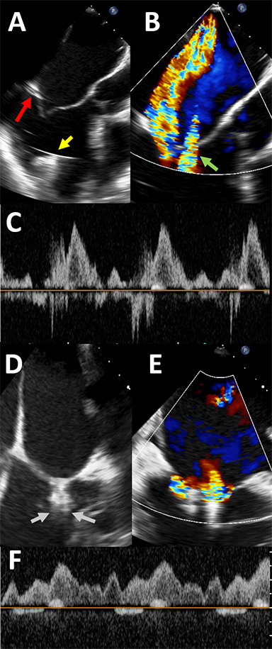
Figure 2.

Intraprocedural transesophageal echocardiogram images for patient 2. Panel A shows tethering of the posterior leaflet (red arrow) as well as the left ventricular assist device (LVAD) inflow cannula (yellow arrow). Panel B shows color Doppler across the mitral valve, confirming significant mitral regurgitation (MR). Nyquist limit is 61 cm/second. “Waterfall artifact” from the LVAD is also seen (green arrow). After one MitraClip XTR is placed, significant residual MR is seen lateral to the clip in the bicommissural view, as seen in panel C, with the MR jet simultaneously visualized in the 3-chamber view. A second MitraClip XTR is then placed just lateral to the first clip, as shown in panel D (gray arrows). Panel E shows color Doppler across the clips, confirming improvement in MR. There is no longer reversal in the pulmonary veins (Panel F).
