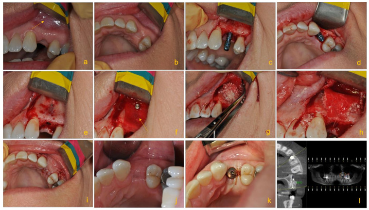
Figure 3.
Clinical images illustrating the pre- and post-operative (post-op) procedures of an edentulous patient treated with an implant with guided bone tissue regeneration. Images from left to right show: (a) the edentulous site after the tooth was extracted; bone defect in the form of buccal concavity is visible in the apical aspect of 24 (yellow arrow). (b) Occlusal shot of the edentulous site showing the buccal concavity. (c) The correct positioning of the implant (Straumann Bone Level Tapered 3.3 × 10 mm implant). (d) Proper bucco-palatal positioning of the implant. (e) Decorticated area to prior to placement of the bone and complete absence of the buccal bone at the apical part of the implant. (f) The placement of Straumann Flex Membrane fixed and stabilized by tacks (AutoTac by BioHorizons Canada). (g) Applying bone graft particles comprising of a mixture of Allograft and Xenograft (both from Straumann®) packed at the buccal bone defect. (h) Periosteal sutures used to stabilize and fix the bone graft inside the membrane, which ensures immobilization of graft resulting in optimal bone regeneration vs. fibrous tissue formation. (i) Primary closure of the site. (j) Showing post-op. Primary closure is intact. (k) The implant after second stage and osseointegration check. (l) Five months later, post-op cone beam computed tomography (CBCT) illustrating the final bone healing prior to second stage and osseointegration check of the implant. The post-op CBCT revealing a gain of over 5 mm of bone (courtesy of Dr. Mohammad A. Javaid, Periodontist, British Columbia, Canada).

