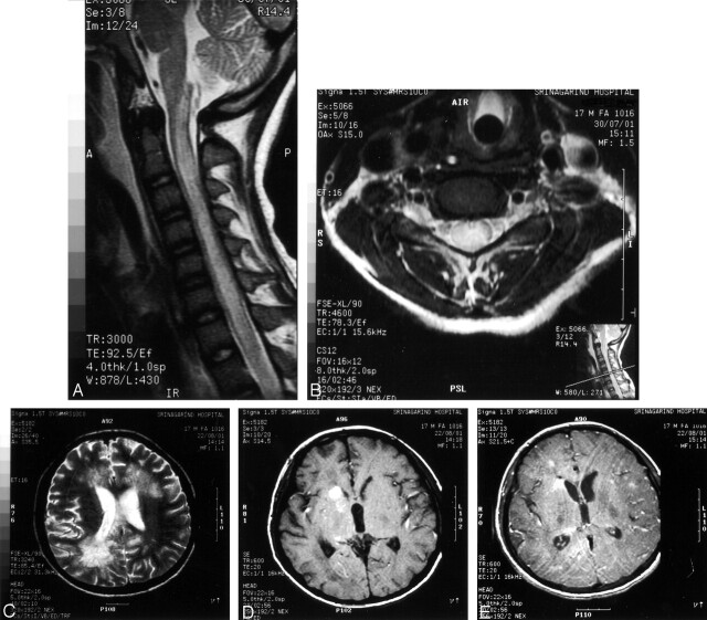
Fig 2.
Case 2. MR images of cervical cord and brain.
A, Sagittal T2-weighted image, showing diffuse cord enlargement with ill-defined area of increased signal intensity.
B, Axial T2-weighted image, showing hyperintense lesion within central gray matter.
C, Axial T2-weighted image, showing fuzzy hyperintense lesion at both periventricular white matter regions.
D, Axial T1-weighted image, showing intracerebral hemorrhage at right caudate nucleus and posterior part of basal ganglia.
E, Axial T1-weighted postgadolinium image, showing scattered tiny nodular enhancement at both frontoparietal regions.

