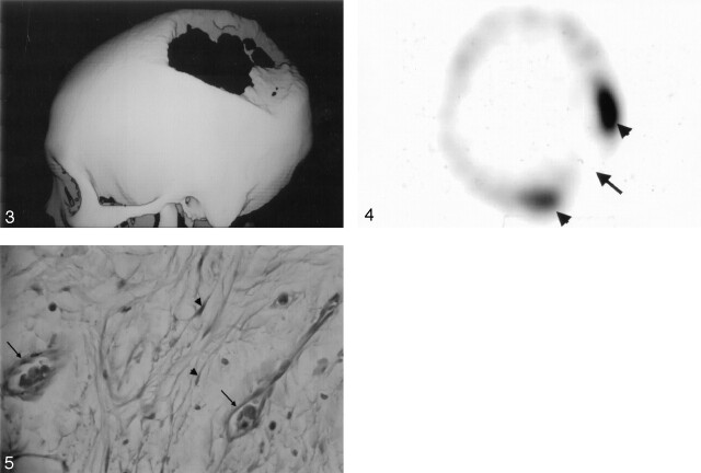
Fig 3.
3D reconstructed CT scan obtained by using a bone algorithm viewed from the left lateral delineates well the large skull defect.
Fig 4. Tc-99 m MDP bone scintigraphy reveals no bone uptake in the resorbed site (arrow) but increased uptake around the margin (arrowheads).
Fig 5. Photomicrograph of the bone specimen shows the small capillary-like vessels (arrows) and spindle-shaped fibroblasts (arrowheads) (hematoxylin and eosin stain, × 400)

