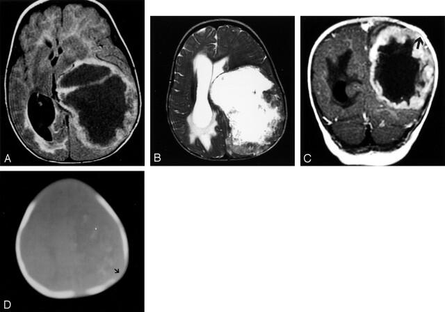
Fig 1.
Patient 1, with lesion in left cerebral hemisphere.
A, Axial view fluid-attenuated inversion recovery image shows a very large cyst mass with thick nodular walls and mass effect but little edema.
B, T2-weighted fast spin-echo image, obtained at the same level, shows a necrotic mass with a thick heterogeneous wall causing midline shift.
C, Coronal view contrast-enhanced image shows a nodular rind of tumor. Remodeling of the calvarium by the mass can be seen (arrow).
D, CT bone window shows erosion of the left parietal bone (arrow) and faint calcification.

