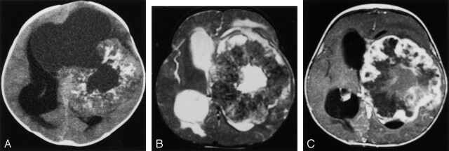
Fig 2.
Patient with left hemispheric mass.
A, Unenhanced CT scan displays a large doughnut-shaped mass with central cyst that opens into an eccentric large cystic component. Lesion has mass effect but little edema. Several calcifications are present in the tumor.
B, T2-weighted fast spin-echo image shows a large thick walled mass with mild surrounding edema. Note that the tumor is hypointense, presumably secondary to the calcifications.
C, Contrast-enhanced T1-weighted axial view image reveals a nodular peripheral enhancement in the thick calcified rind.

