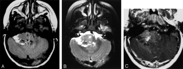
Fig 4.
Cerebellopontile angle mass.
A, Fluid-attenuated inversion recovery image shows a right cerebellopontile angle extra-axial mass with solid and peripheral cystic components. Focal hypointensities correspond to calcifications shown on CT scans (not shown).
B, T2-weighted fast spin-echo image shows a right cerebellopontile angle extra-axial mass with solid and peripheral cystic components. Focal hypointensities correspond to calcifications shown on CT scans (not shown).
C, Contrast-enhanced image shows mild enhancement with a small, relatively more enhancing nodule.

