
Fig 2.
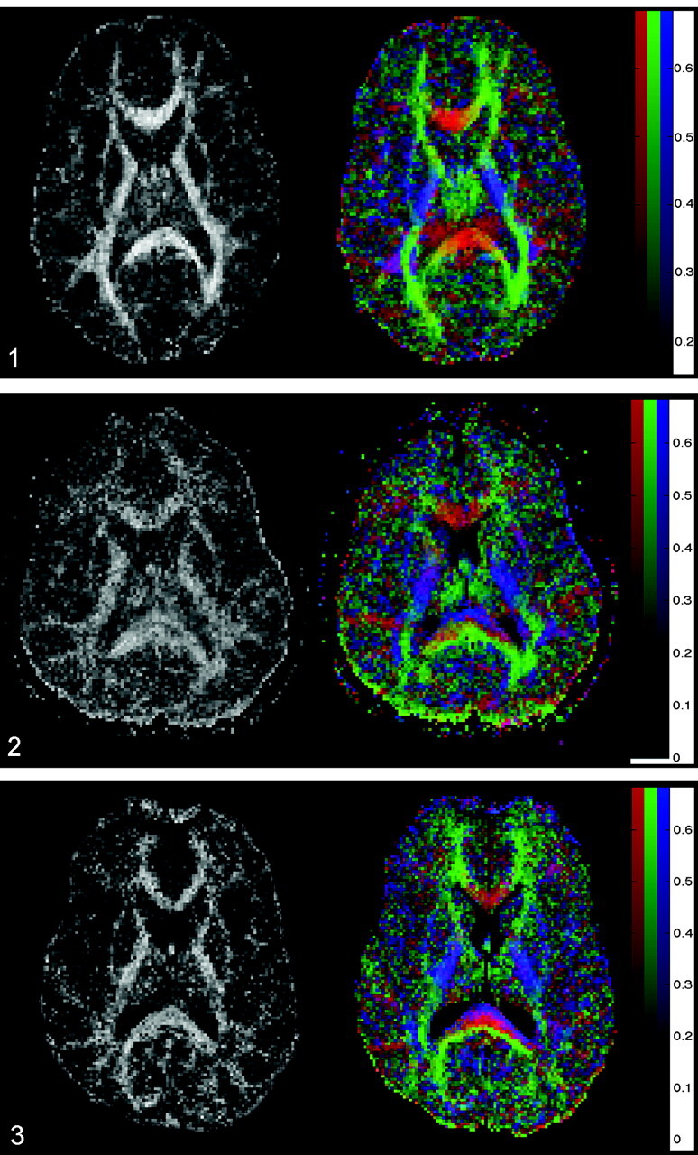
Images in a 24-year-old man with severe TBI. Acute GCS, 5. Rankin score at discharge, 3. Left, FA map shows a reduced FA index of the splenium of the corpus callosum (FA = 0.511 ± 0.036, mean control FA = 0.808 ± 0.060) and internal capsule (FA = 0.531 ± 0.036, mean control FA = 0.735 ± 0.066). Right, Color-coded map shows that, within the center of the splenium of the corpus callosum, the normally predominant red voxels are missing and replaced by a mixture of blue and green voxels (compare with Fig 1). This finding suggests that fiber tracts that connect both cerebral hemispheres are injured or disrupted within the center of the splenium.