
Fig 1.
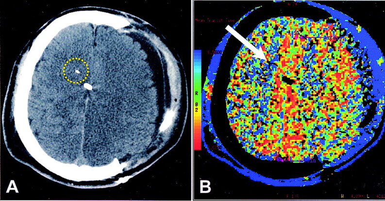
Patient who underwent hemicraniectomy after head trauma.
A, Nonenhanced CT scan shows the tip of the brain-tissue oxygen probe as a white linear attenuation in the right hemispheric white matter. Midline white attenuation is a ventriculostomy catheter. Yellow circle indicates the ROI for CTP analysis.
B, CTP color map of MTT. On the reference bar, blue indicates the slowest MTT. Arrow points to the tip of the oxygen probe. Overall, MTT is decreased on the side of hemicraniectomy, indicating more rapid transit. Surrounding blue rim represents scalp perfusion.