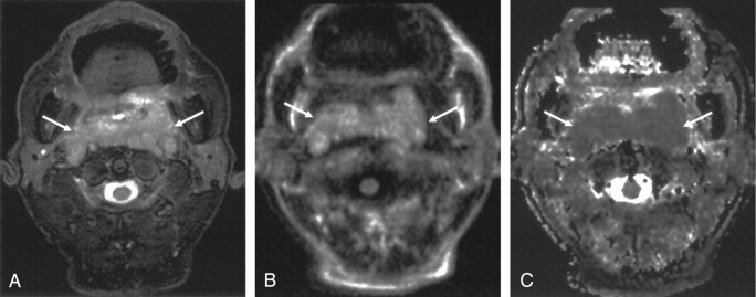
Fig 2.
Axial images obtained in a 69-year-old man with diffuse large B-cell lymphoma of the nasopharynx.
A, T2-weighted fat-suppressed MR image shows the mass (arrows).
B, LSDWI obtained with b = 1000 s/mm2 shows the mass (arrows).
C, Corresponding ADC map. ADC value of the mass (arrows) is 0.69 × 10⁻³ mm2/s.

