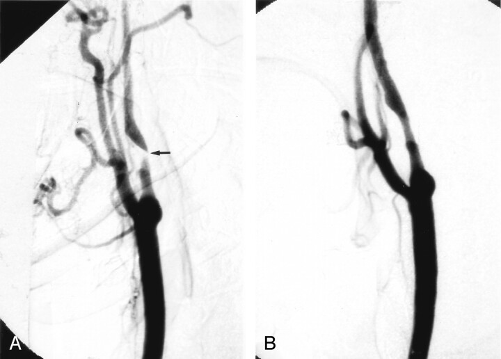
Fig 1.
A, Right CCA digital subtraction arteriogram (DSA), lateral view, showing a very severe atherosclerotic stenosis of the proximal ICA, >95% by NASCET criteria (arrow).
B, Repeat DSA, lateral view, immediately poststenting alone, without balloon angioplasty, showing a reduction in the degree of stenosis to approximately 29%.

