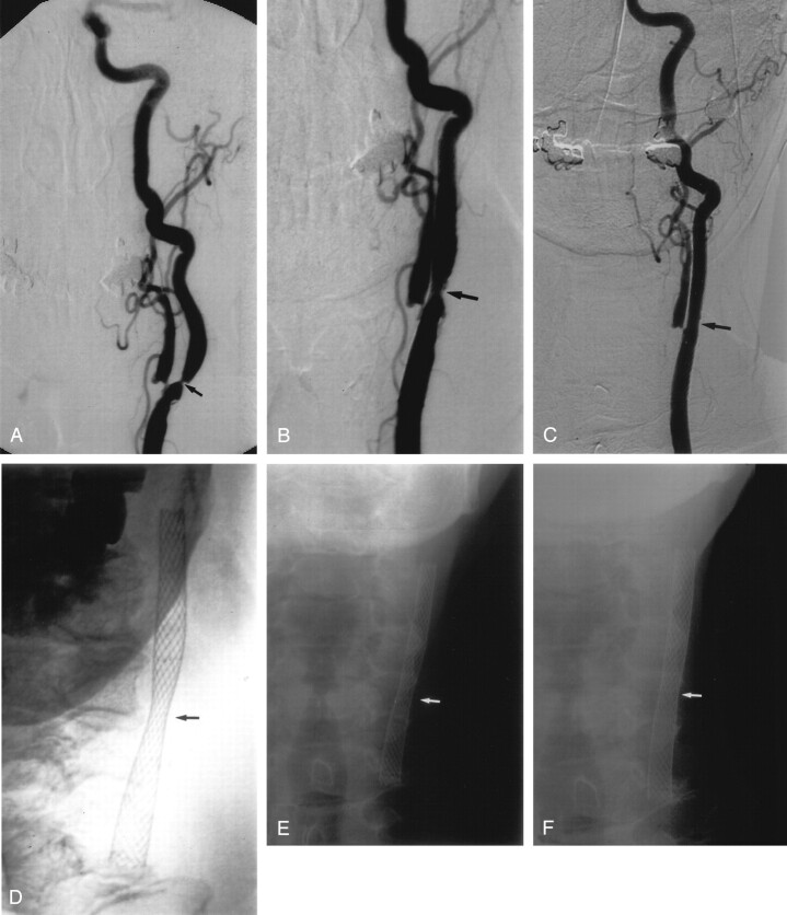
Fig 2.
A, Left CCA DSA, AP view, showing a severe stenosis of the proximal ICA, measuring approximately 76% (arrow).
B, Repeat DSA, AP view, immediately poststenting without balloon angioplasty, showing reduction of the stenosis to approximately 50% (arrow).
C, Follow-up DSA, AP view, 3 years poststenting alone, shows no residual ICA stenosis.
D, E, and F, Conventional AP radiographs of the neck immediately poststenting (D), 1 month (E) and 8 months (F) poststenting, showing progressive opening of the stent waist (arrow), with maximum expansion occurring in the 1st month postprocedure.

