
Fig 3.
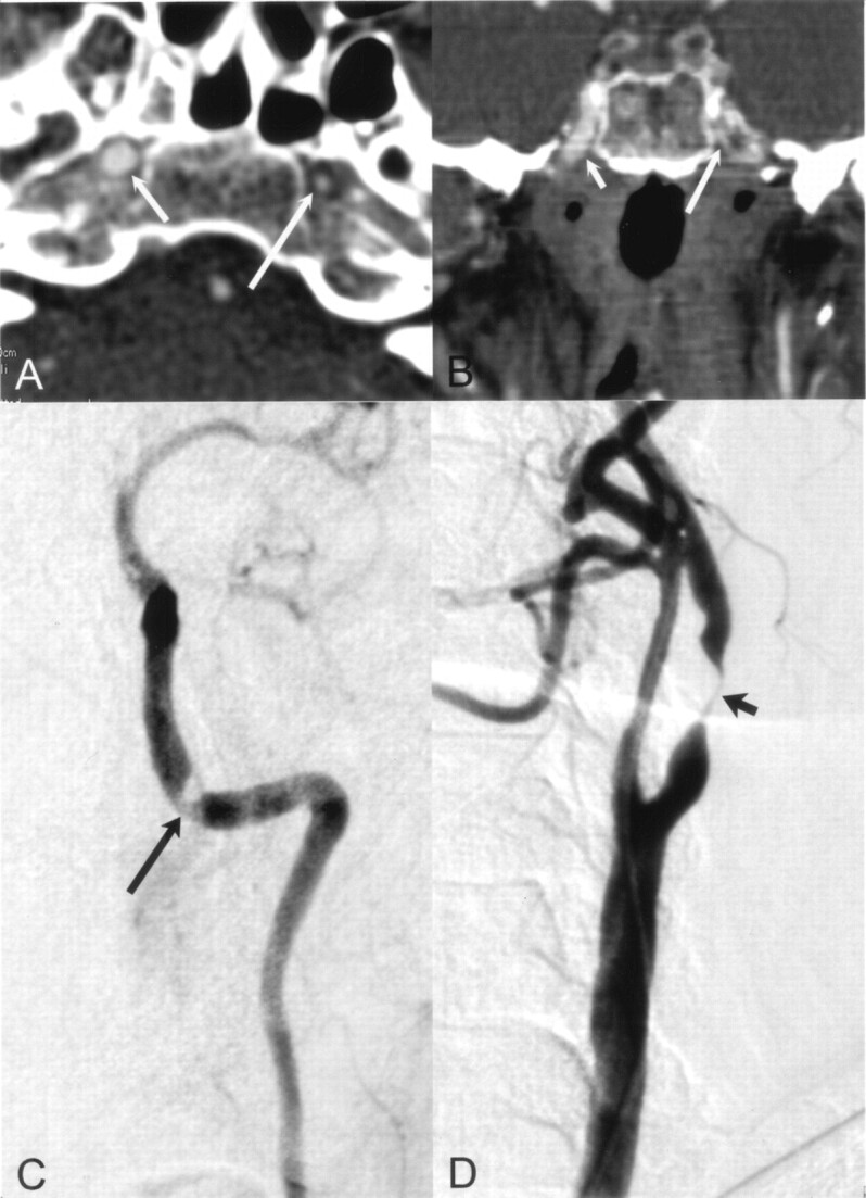
Distal lesion in the petrous part of the left ICA detected correctly with CT angiography.
A and B, Axial (A) and coronal (B) MPR images of distal ICAs show the smaller diameter of the left ICA (long arrow) compared with the normal ICA on the right (short arrow).
C and D, Selective angiograms verify the stenosis (arrow in C) and a near-occlusion (arrow in D) at the bulb. This tandem lesion on the symptomatic side was successfully treated with angioplasty and stent placement.