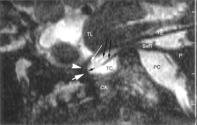
Fig 2.
Sagittal nonenhanced 3D CISS image through the right side of the Meckel cave in a 58-year-old woman. Sensory root (SeR) is shown from its apparent origin at the pons (P) to the entrance of the cave. Ganglion (thick white arrow) at the anterior aspect of the cave is difficult to identify, and anterior margin of the trigeminal ganglion cannot be distinguished from the dural wall (arrowhead) of the cave. Only the superior lip of the ganglion (thin white arrow) is well defined. One smaller and one larger sensory rootlet (long black arrows) arise from the sinus ganglii (short black arrow) and course in the trigeminal cistern (TC). Maxillary nerve is not depicted at the inferior border of the cavernous sinus. CA = carotid artery, PC = prepontine cistern, TE = cerebellar tentorium, TL = temporal lobe.

