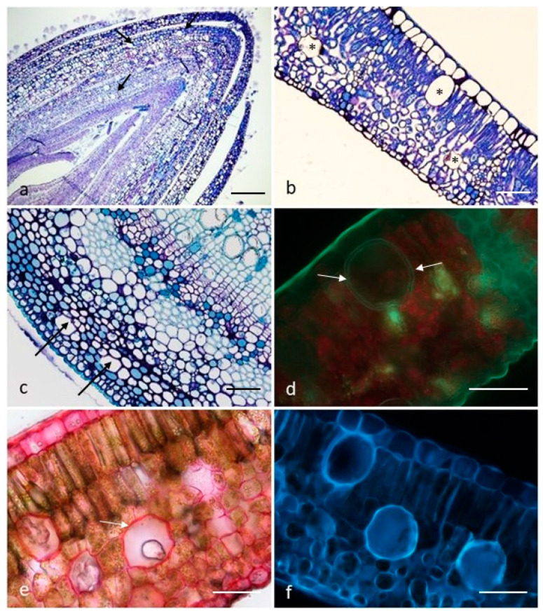
Figure 1.
(a–c). Transverse sections of a leaf bud (a), a full-expanded leaf (b) and a young stem (c) showing the distribution pattern of the secretory cells (arrows and asterisks). Toluidine Blue. (d–f). Transverse sections of full-expanded leaves: primary fluorescence under UV light (d), notice the two-layered cell wall (arrows); Ruthenium Red test on the secretory cell walls (arrows) (e); primary fluorescence under Blue light (f). Scale bars = 200 µm (a); 100 µm (c); 50 µm (b,d–f).

