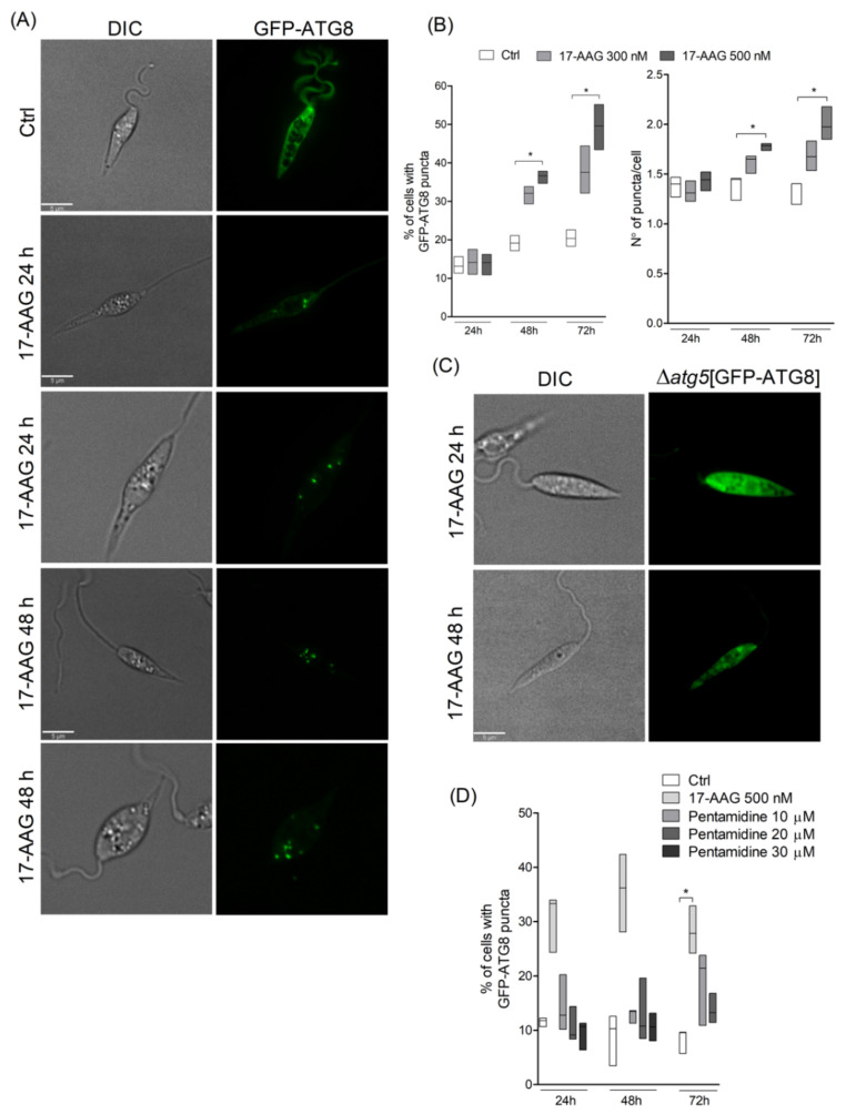
Figure 1.
Evaluation of autophagosome formation in Leishmania promastigotes following treatment with 17-AAG. (A) Axenic promastigotes of Leishmania expressing GFP-ATG8 were treated or not with 17-AAG (500 nM) for 24 or 48 h and imaged by fluorescence microscopy. (B) The percentage of cells bearing autophagosomes and the number of autophagosomes per cell were calculated at 24, 48 and 72 h after treatment with 17-AAG (300 or 500 nM). (C) Δatg5[GFP-ATG8] parasites were treated with 17-AAG and imaged by fluorescence microscopy. (D) Comparison of the percentage of cells bearing autophagosomes after treatment with pentamidine (10, 20 or 30 μM) or 17-AAG (500 nM) for 24 and 48 h. Lines within the floating bars represent medians and floating bar quartiles (Q: 25% and 75%) from one out of three independent experiments (Kruskal-Wallis test, Dunn’s multiple comparison test, * p < 0.05).

