Figure 2.
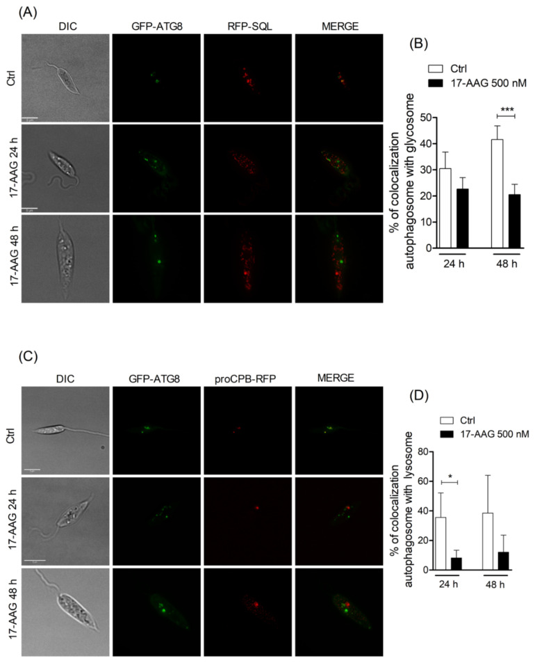
Analysis of fusion between autophagosomes and glycosomes or lysosomes. (A) Axenic promastigotes of Leishmania expressing GFP-ATG8 and RFP-SQL were treated or not with 17-AAG (500 nM) and imaged by fluorescence microscopy. (B) Quantification of autophagosome-glycosome colocalization after treatment of Leishmania with 17-AAG. (C) Axenic promastigotes of Leishmania expressing ATG8-GFP and proCPB-RFP were treated or not with 17-AAG (500 nM) and imaged by fluorescence microscopy. (D) Quantification of Leishmania autophagosome-lysosome colocalization after treatment with 17-AAG. Bars represent medians ± SD from one out of three independent experiments (Unpaired t test, *** p = 0.0006, * p = 0.0197).

