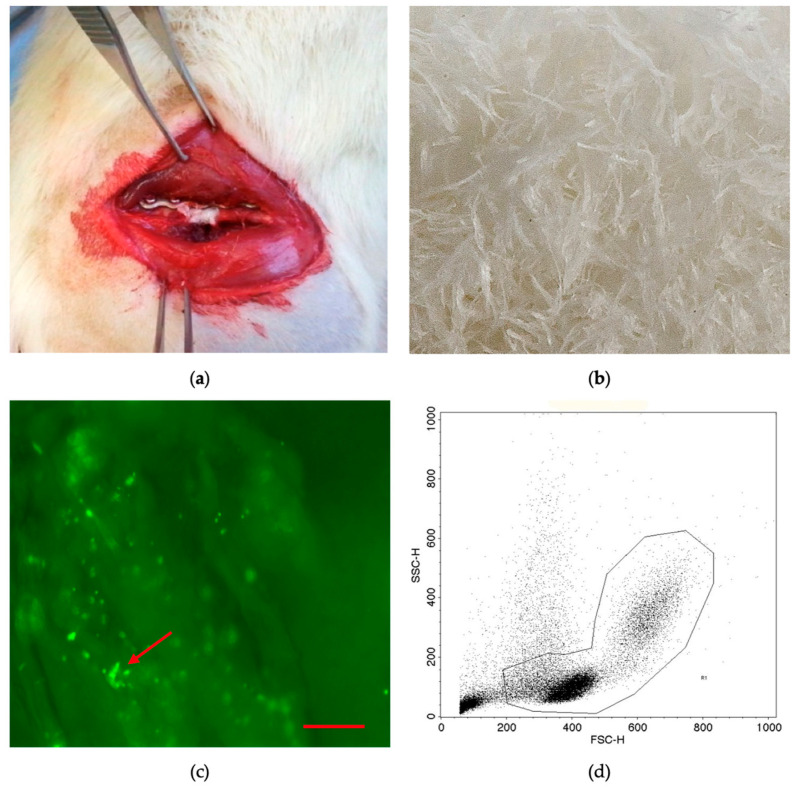
Figure 1.
Plate-stabilized and filled femoral defect (a). A 5 mm critical-size defect in the right femur of a rat is stabilized with a 5-hole plate. The gap is filled with demineralized bone matrix fibers (f-DBM) loaded with BMCs. Macroscopic image of the fibrous tissue scaffold (b). Fluorescence microscopic image; adherent CFSE-stained BMCs (green dots, marked with red arrow) can be seen at different depth levels of the f-DBM plexus (original magnification 50×) (c). FACS characterization of rat BMCs (d). Mainly mononuclear cell types (lymphoid and monocytic cells) remain after density gradient centrifugation.

