
Figure 10.
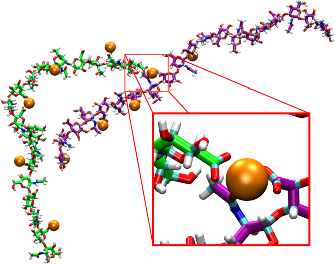
Intermolecular association between O-type chondroitin polymer strands bridged by Ca2+. The two chondroitin polymer strands are colored green and purple, respectively, and bound Ca2+ cations are shown as orange spheres. The conformation is from a simulation with the c36_jul17 force field (i.e., O’Cal).