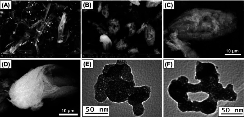
Figure 2.
SEM image of diatomite precursor (A), synthetic CS/D composite (B), high-magnification image of the chitosan cluster on the surface of diatomite frustules (C), high-magnification image of the formation of chitosan as nanofibers (D), and TEM images of the CS/D composite reflecting enclosing of diatomite grains within the chitosan matrix (E, F).

