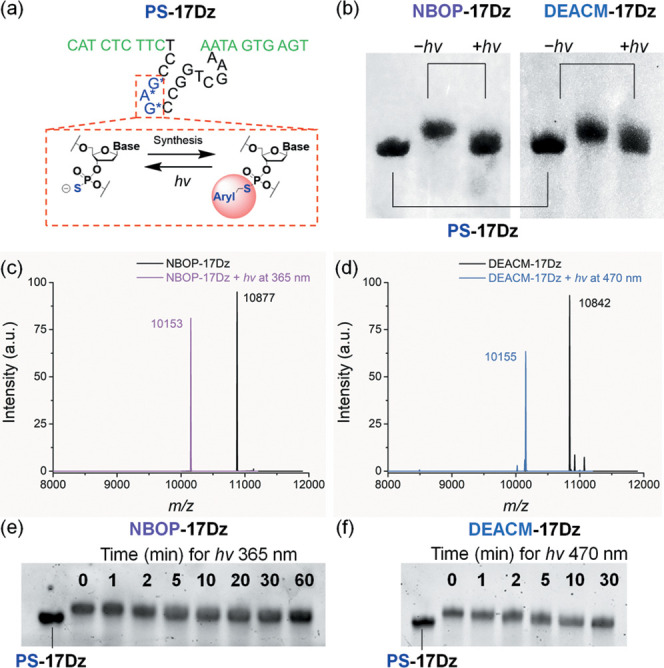
Figure 2.

(a) Preparation of photocaged DNAzymes through the postsynthetic derivatization of PS and light-induced decaging. The binding arms of the DNAzyme are colored in green and the catalytic core is colored in black. The blue G*A*G* region contains PS modifications in their phosphorodiester linkages. For NBOP-17Dz, Aryl = NBOP; for DEACM-17Dz, Aryl = DEACM. All three PS sites are subjected to modification. (b) PAGE analyses of PS-17Dz, NBOP-17Dz, and DEACM-17Dz treated with or without light. (c) ESI-MS analyses of NBOP-17Dz before (m/z, found 10 877, NBOP-17Dz calcd 10 876) and after (m/z, found 10 153, PS-17Dz calcd 10 153) light activation. (d) ESI-MS analyses of DEACM-17Dz before (m/z, found 10 842, DEACM-17Dz calcd 10 840) and after (m/z, found 10 155, PS-17Dz calcd 10 153) light activation. (e) Time-dependent decaging of NBOP-17Dz by 365 nm light irradiation. (f) Time-dependent decaging of DEACM-17Dz by 470 nm light irradiation. For all analyses, 10 μM DNAzyme was prepared in sodium phosphate buffer (pH 7.0). Power of light irradiation: 365 nm at 26 mW/cm2 and 470 nm at 13 mW/cm2.
