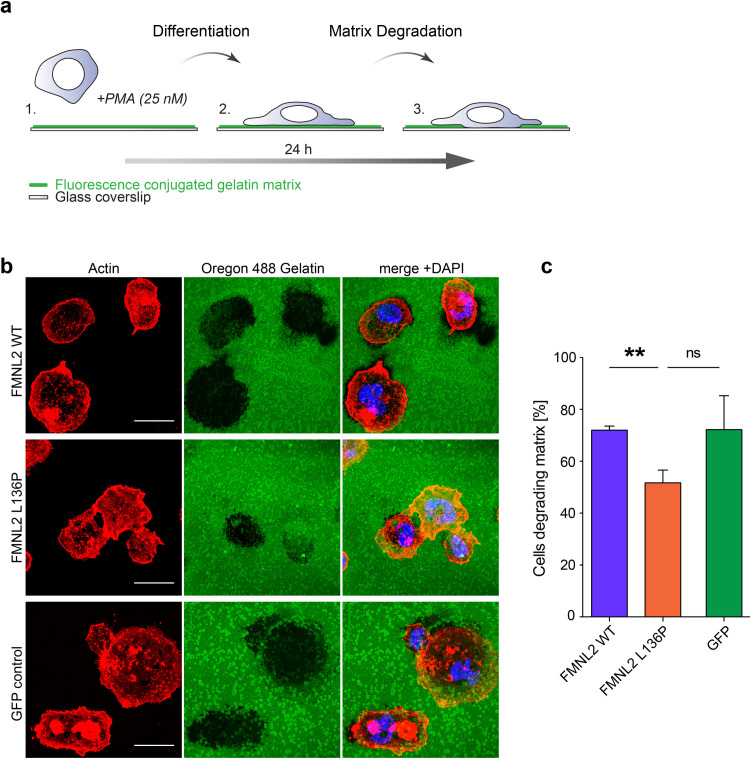
Fig 5. The formin mutant disturbs matrix degradation in THP-1 macrophages.
a) Schematic cartoon of the matrix degradation assay. THP-1 cells were seeded on fluorescence labeled gelatin matrix with PMA treatment over 24 hours. PMA stimulates the differentiation from monocyte into macrophage state. Dynamic podosome activity enables macrophages to degrade the matrix over time via enzymatic digestion. b) Matrix degradation of THP-1 macrophages. THP-1 cells were seeded on glass coverslips coated with fluorescence-labeled gelatin matrix. After fixation cells were stained with Alexa Phalloidin to visualize actin structures. Digested matrix appears black in the green gelatin matrix layer. Scale bar = 20 μm. c) Quantification of cells showing degradation activity (black areas in the fluorescent matrix) was performed on tile scan images representing > 100 cells per experiment. Data are shown as mean (+SEM) of 3 independent experiments (L136P vs. WT: p = 0,002; L136P vs. GFP: p = 0,064). Students T-test was used to analyze the statistical difference between two groups (* = p<0,05; ** = p<0,01).

